
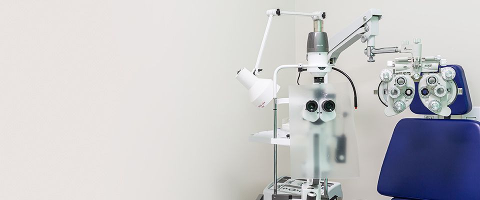
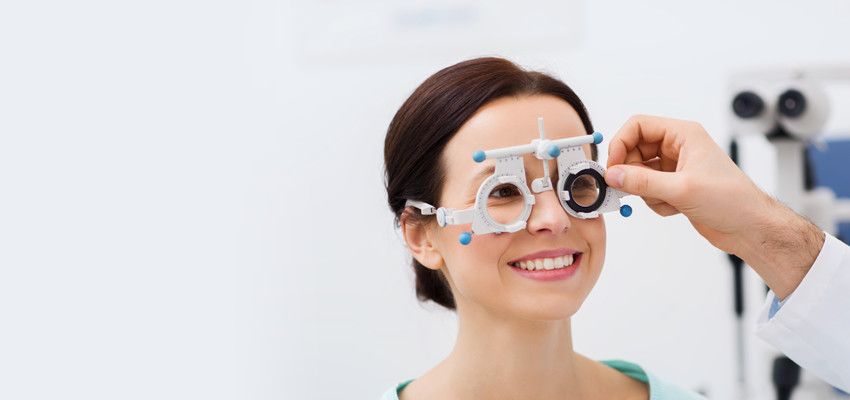
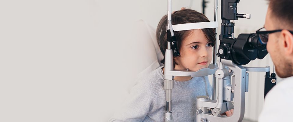

Meet Our Doctor
Our doctors are leaders in eye care offering optical services that provide the best eye care solutions to all our patients.
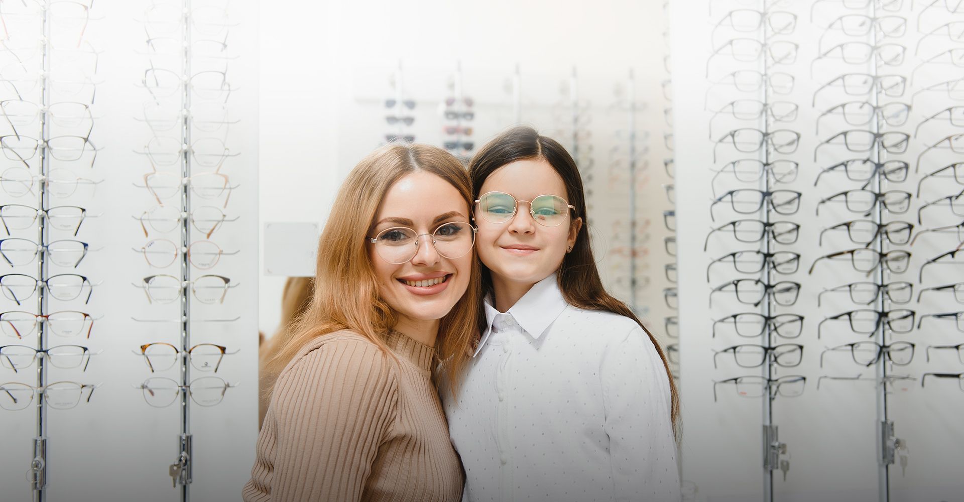
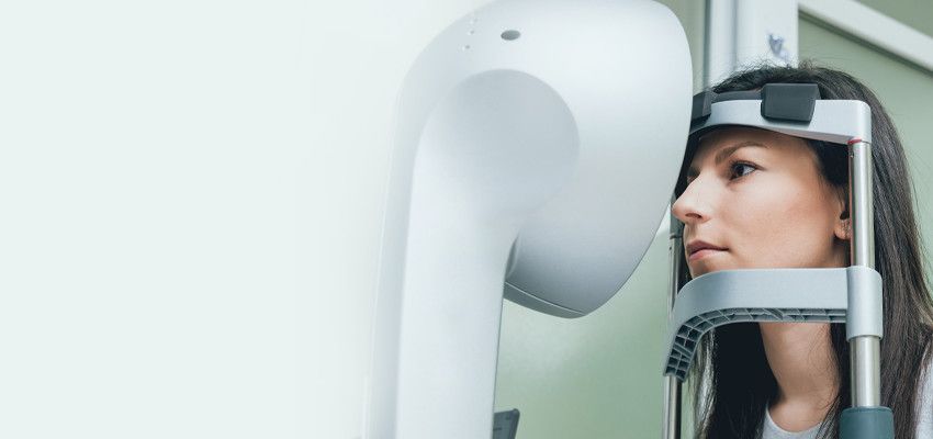
We're a sincere company with a straightforward vision. With us, vision care goes far beyond a simple eye examination.
Our doctors are leaders in eye care offering optical services that provide the best eye care solutions to all our patients.
© 2024 Mead EyeCare & EyeWear. All rights Reserved. Accessibility Statement - Privacy Policy - Sitemap
Powered by: